Researchers are using an advanced imaging technique to help diagnose mild blast-related traumatic brain injuries in soldiers whose wounds don’t usually show up in conventional brain imaging scans. Scientists are hopeful the new technique will enable doctors to identify many previously untreated brain-injury victims, and help them get the medical care they need.
An estimated 320,000 military personnel in Iraq and Afghanistan have suffered traumatic brain injuries. Often these injuries are classified as mild because they do not show up on brain scans, making subsequent symptoms, like brain fog or forgetfulness, difficult to treat.
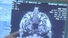
But in a new study, an advanced brain imaging technique called diffusion tensor imaging was able to find abnormalities in the white matter brain tissue of soldiers who had sustained mild blast-related traumatic brain injuries, or TBI. White matter is a critical part of the brain’s wiring system that allows nerve cells to communicate with one another.
Conducted by investigators at the Washington University School of Medicine in St. Louis and the Landstuhl Regional Medical Center in Germany, the small study involved 63 soldiers who had sustained mild TBIs as a result of battlefield blasts plus other injuries such as falls, motor vehicle injuries or being struck by blunt objects.
There was also a control group of 21 military personnel who had been injured in blasts but were not diagnosed with TBI.
Washington University neurology researcher Christine MacDonald says tensor imaging allows scientists to see the movement of water through injured brain tissue, something that’s not seen in other types of brain imaging.
MacDonald says investigators noted the presence of water in the white matter on initial scans taken in Germany approximately fourteen days after the initial injury.
MacDonald says researchers then saw faint but discernable changes in water flow, or diffusion, in follow-up tensor scans six to 12 months later, after the soldiers had returned to St. Louis. "This diffusion of water is in line with what we expect for injury and its progression over time. Versus something that would, say, be pre-existing," she said.
Researchers noted white matter abnormalities in the tensor images of 18 of 63 patients in the study who had been diagnosed with concussions. They were not seen in 21 patients diagnosed with other types of injuries.
A diagnosis of TBI is controversial because experts are not sure whether the symptoms following mild traumatic brain injury are due to disruptions in brain chemistry, structural changes, physiological factors or a combination of all three.
At this point, MacDonald says experts are unsure what role, if any, the white matter abnormalities play in factors like altered attention, memory and, importantly, post-traumatic stress disorder, a condition which plagues many returning veterans.
MacDonald says studies continue to explore whether diffusion imaging could be a useful tool in helping to identify the source of the lingering effects of traumatic brain injury.
“Long term, years down the line, possibly this could be something that would be an assisted diagnostic tool. But that’s really something that’s outside the scope of this current manuscript; but definitely hope long term,"she said.
An article describing the discovery brain abnormalities in people with mild blast-related traumatic brain injury is published in the New England Journal of Medicine.