The world's first microscope that can see active infectious diseases in super-resolution video has been installed at a university in Australia. The technology promises new insights into malaria and viruses that cause potentially fatal infections. The Microbial Imaging Facility is capable of producing real-time, multiple color images of diseases attacking living cells. Researchers say it has the potential to help develop treatments for a range of life-threatening conditions.
The super resolution microscope takes up an entire room at the University of Technology, Sydney, where the air is filtered to prevent dust from damaging the sensitive equipment.
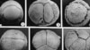
The speed of the microscope's cameras and computers allows researchers to see diseases like never before.
Lynn Turnbull is in charge of its operation. “At one end we have the ability to see the resolution down to 100 nano meters, which is a tenth of the size of a bacteria. So you’re looking inside bacteria now instead of just looking at blurry little blobs on a screen,” she explained.
The technology is called the DeltaVision OMX Blaze super resolution imaging system. It was developed in the United States and costs about $1 million. Turnball says the microscope gives live images of infectious diseases as they infiltrate healthy cells. “Though we can’t capture things that move in less than a second, now we’re actually in the realms of watching processes inside cells, watching bacteria invade cells or malaria parasites invade a cell that we can now start to look at these processes in real time instead of just taking snapshots,” she said.
Scientists are now seeing bacteria, parasites and viruses in unprecedented detail.
Professor Ian Charles from the University of Technology, Sydney says the technology could give medical science a decisive advantage in the fight against disease.
“We’re actually beginning to see sub-structures of the micro-organism that we hadn’t seen before, not only in terms of the bug itself but the way the bug interacts with its host cell and does whatever it needs to do to get into the cell and get out of the cell," explained Charles. "So I think it can be a real beginning of a formulation of perhaps a new way to look at what the micro-organisms are actually doing.”
The team in Sydney is also able to share its microscope with other institutions around the world through a video-sharing network.
Professor Charles says images are fed from the microscope to a large bank of computer screens that can be linked to universities and hospitals in other countries.
“This is the device that allows us to look in real time in groups of scientists who are interested in doing the experiments, and actually help direct the experiments actually on the microscope," Charles said. "So we can sit around here discussing the experiments, looking at the screen and actually working out where to look next.”
So there could be someone in North America or someone in Europe looking at this screen in the same time?
"In real time looking at the way their experiments are being run,” Charles stated.
Researchers hope the technology here in Sydney will help in the development of new drugs by giving scientists the opportunity to better understand how microorganisms such as malaria cause infection.