

Three-D printing technology is now being applied to the human heart in order to help surgeons better assess patients before going under the knife.
While surgeons routinely use digital images to explore the heart in minute detail before conducting an operation, no two human hearts are exactly alike. This fact has led Dr. Matthew Bramlet to begin conducting more comprehensive research on the 3-D heart models that can be produced from those images.
As co-author of a study on medical possibilities made available via 3-D printing, he recently presented his findings at the annual American Heart Association meeting in Chicago.
“The detail that we’re able to achieve is just remarkable,” he told conference goers, holding aloft a 3-D printed baby heart about the size of an egg. “It still baffles my mind that the surgeons are able to do the things that they are able to do.”
The University of Illinois College of Medicine pediatric cardiologist — Bramlet is not himself a surgeon — says the 3-D replica uncovers information that can’t be obtained any other way.
“Even when I take the MRI and render the images — even a print heart that’s printing, that is spinning on the screen — it’s still a 2-D screen,” he said, explaining that 3-D printing allows him to virtually grab those images off the screen.
"You can actually hold it in your hands and evaluate the anatomy for the first time,” he said.
Here's how it works: A C-T scan, MRI and other data are sent to a 3-D printer to replicate a heart in a plaster or ceramic mold. The printer then constructs the heart, thin layer by thin layer. Bramlet says the model matches the real heart with an unprecedented level of accuracy and detail.
“When we’re done with the [printed] model and made our decision, we want to be able to go back to the source image and confirm those findings,” he added.
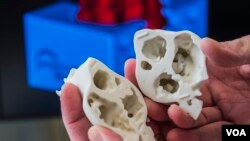
Bramlet has built model hearts for various procedures, all with successful outcomes. In his first case, digital images showed one tiny hole in a baby’s heart, yet the 3-D printed model revealed multiple holes — birth defects not readily apparent in the standard images.
“Knowing that it was there, we could still not demonstrate it [in the images], and, sure enough, [the surgeon was] able to confirm it and change the surgery," he said. "In the future, relying on that information would allow us to not even have to stop the heart and to go down an alternative pathway.”
Bramlet's research follows numerous accounts of surgeons successfully using 3-D printing technology before heading to the operating table. Dr. Emile Bacha, head of cardiac surgery at Morgan Stanley Children's Hospital of New York Presbyterian, reported success with a similar approach to preparing surgery for a 2-week-old infant in July. According to reports in American and British news outlets, Bacha received funding from Matthew's Hearts of Hope to used Materialise's Mimics Innovation software to create a replica of the baby's heart, which was "riddled with holes and structured unusually."
Like Bramlet, Bacha described the 3-D replica as a roadmap to guide his surgical team, such that they were able to fix the baby's heart with a single procedure as opposed to conducting a series of operations.
According to Kathy Magliato, a cardiac surgeon at Saint John’s Health Center in Los Angeles, the new technology is a potential game-changer for cardiac surgeons everywhere.
“The fact that I can then take this very complicated structure, which has endless possibilities of what it could look like anatomically when I open the body, and you give it to me before the operation and I can hold it in my hand and plan an operation around what I’m seeing ... that to me is what can potentially change the game in an operation and save lives.”
Bramlet recently partnered with the National Institutes of Health to build a 3-D reference library that includes models and images that others can learn from. The collection will be hosted by the Jump Trading Simulation and Education Center in Peoria, Illinois.